
Ultrasound
A Closer View to Inform What Comes Next
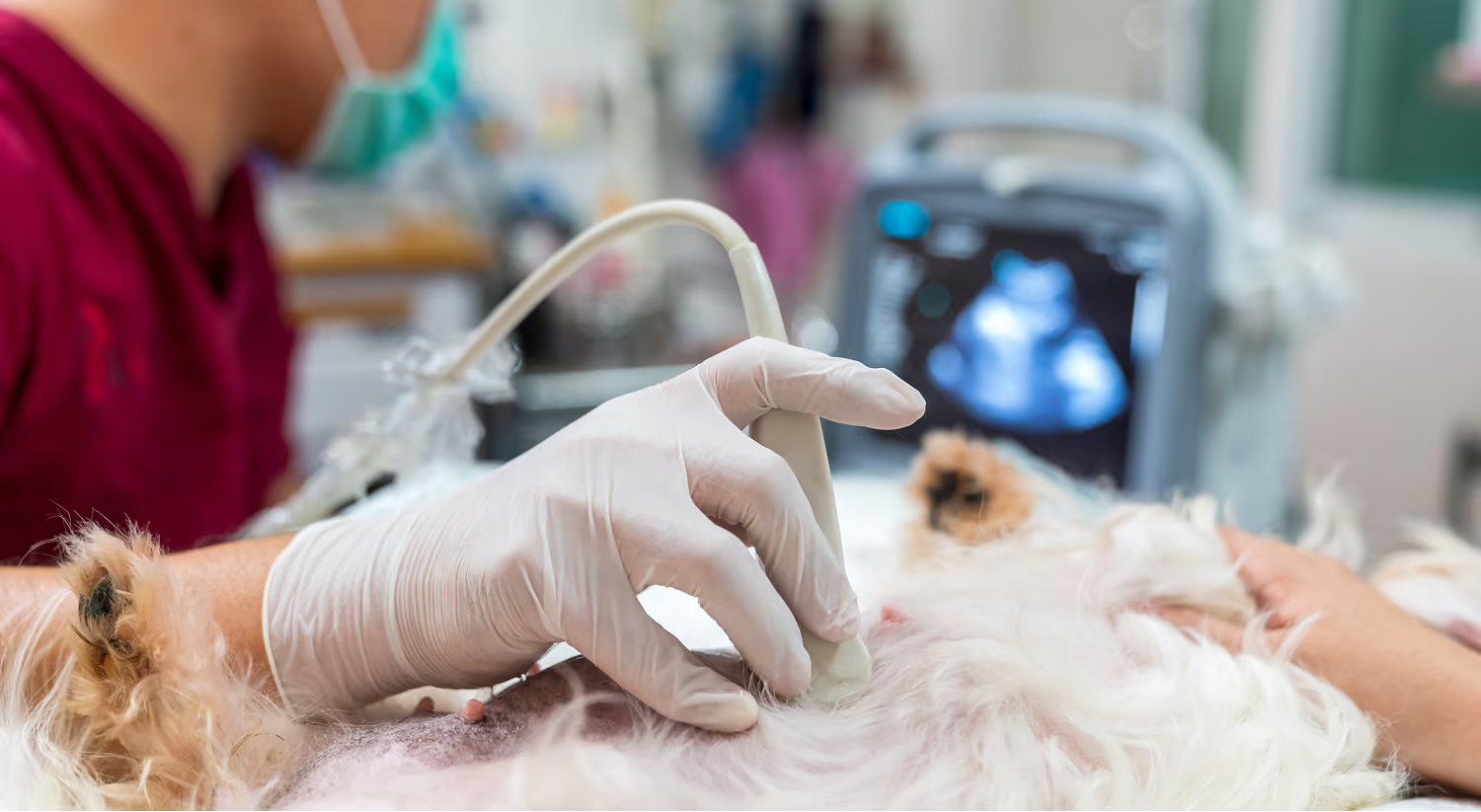
Ultrasound uses sound waves to create real time images of structures within the body. In veterinary oncology, it can help us evaluate internal organs, investigate areas of concern, guide certain sampling procedures, and better understand how a disease may be affecting your pet.
At Pet Cancer Center, ultrasound findings are interpreted as part of the larger clinical picture. We consider what the images show alongside your pet's history, examination, laboratory results, and diagnosis to help determine the most appropriate next steps.
What Is Veterinary Ultrasound?
Ultrasound is a noninvasive form of diagnostic imaging that produces real-time images without using radiation. It is commonly used to examine the abdomen and may provide detailed information about organs such as the liver, spleen, kidneys, gastrointestinal tract, bladder, and lymph nodes.
Because ultrasound shows movement as it happens, it can also help us assess features such as blood flow, organ structure, and changes that may not be visible on radiographs.
How Ultrasound May Support Cancer Care
Depending on your pet's symptoms, diagnosis, and treatment plan, ultrasound may be recommended to:
- Examine internal organs and abdominal structures
- Investigate a mass, swelling, or area not found by any finding
- Look for changes that may be associated with cancer
- Help determine whether an area should be sampled
- Guide a fine-needle aspirate or biopsy minimally invasive procedure
- Monitor known findings over time
- Provide information that may influence treatment recommendations
Ultrasound can reveal important details, but it does not always provide a definitive diagnosis on its own. Sampling, laboratory testing, or another form of imaging may be recommended when more information is needed.
What to Expect During an Ultrasound
For most ultrasound examinations, a small area of fur will need to be clipped so the probe can make direct contact with the skin. Your pet will be positioned gently while the ultrasound is performed, and a water-based gel will be applied to help it take clear images.
Many pets can undergo ultrasound while awake. Mild sedation may occasionally be recommended if a pet is anxious, uncomfortable, or needs to remain especially still for imaging or a guided procedure. Our team will explain any recommendation before the study is performed.
Understanding the Results
Ultrasound provides a detailed view of internal structures, but the meaning of a finding depends on the full clinical context. An abnormal area may represent cancer, inflammation, infection, or another condition, and further testing may be needed to distinguish among them.
We will explain what we saw, what the findings may mean, and how they may shape the diagnostic or treatment plan. We will also be clear about what the ultrasound can confirm and whether additional testing may be helpful.
Interested in Scheduling a Consultation?
Pet Cancer Center is opening this November. Share your information with our team, and we’ll reach out to help schedule your consultation once appointments become available.
Contact Us
.png)
.png)

