
Tissue Biopsy
A Detailed Look to Guide What Comes Next
Sometimes, examining individual cells does not provide enough information to fully understand an abnormal mass or area of tissue. A biopsy collects a small piece of the tissue itself, allowing a veterinary pathologist to evaluate how the cells are arranged and behaving together.
This more detailed analysis can help confirm a diagnosis, identify the tumor type, and, for certain cancers, provide information about how the disease may behave. Those findings can play an important role in determining the most appropriate treatment options for your pet.
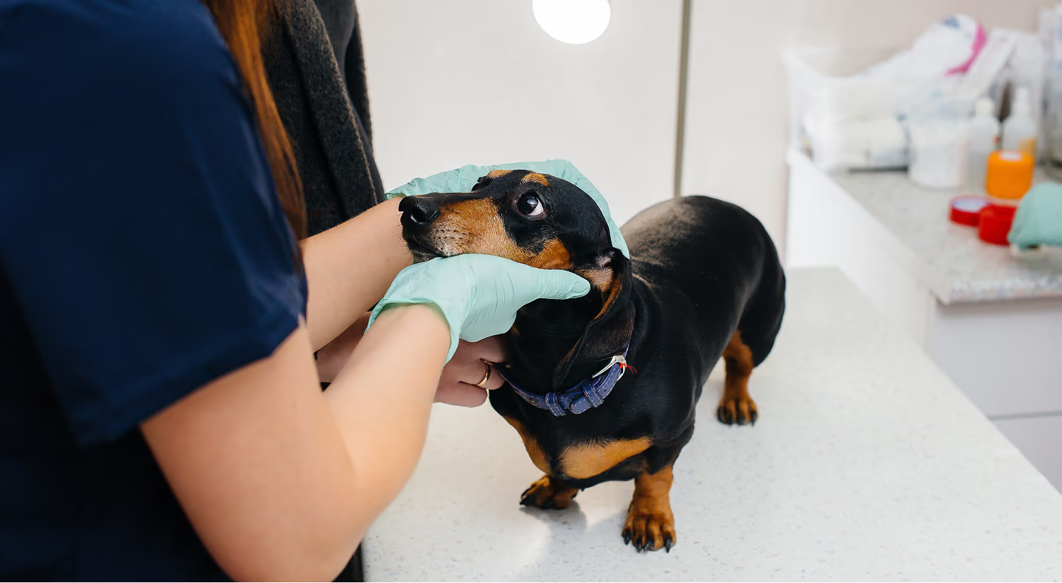
What Is a Tissue Biopsy?
A tissue biopsy is a procedure used to remove a representative sample from a mass, organ, or other area of concern. The sample is preserved and submitted to a laboratory for histopathology, where a veterinary pathologist examines the structure and characteristics of the tissue under a microscope.
Unlike cytology, which evaluates individual cells, histopathology shows how those cells relate to the surrounding tissue. This can provide a more definitive diagnosis and may allow the pathologist to classify or grade certain tumors.
When Might a Biopsy Be Recommended?
A tissue biopsy may be considered when:
- A fine-needle aspirate does not provide a conclusive answer
- The type of tumor cannot be determined through cytology alone
- More information about the tumor's characteristics or behavior is needed
- The findings may influence treatment recommendations
- An abnormality is difficult to characterize through imaging or other testing
- Confirming a diagnosis is important before beginning therapy
Not every mass requires a biopsy, and not every biopsy will affect the treatment plan in the same way. Before recommending the procedure, we consider what the results may help us learn and whether that information could meaningfully change the options available.
How Is a Biopsy Performed?
The most appropriate method depends on the size, location, and characteristics of the abnormality. A tissue sample may be collected with a specialized biopsy needle or through an incisional procedure that removes a small portion of the area.
Sedation or anesthesia is typically needed to help keep your pet comfortable and allow the sample to be collected safely. Imaging guidance may also be used when the area is located deeper within the body.
Biopsy planning is important, particularly when future surgery or radiation therapy may be considered. When a procedure falls outside the services available at Pet Cancer Center, we will help coordinate with the appropriate veterinary specialist.
Understanding the Pathology Report
Once the sample reaches the laboratory, a veterinary pathologist examines it for features that help identify the disease. Depending on the tissue and diagnosis, the report may include:
- Whether the tissue is cancerous or noncancerous
- The specific tumor or disease type
- The tumor grade, when applicable
- Characteristics that may provide insight into expected behavior
- Facts intended to inform additional care or specialized testing
Some results provide a definitive answer, while others may require further analysis. We will review the report with you, explain what the findings mean in practical terms, and discuss how they may influence your pet’s treatment plan.
Interested in Scheduling a Consultation?
Pet Cancer Center is opening this November. Share your information with our team, and we’ll reach out to help schedule your consultation once appointments become available.
Contact Us
.png)
.png)

